
Immunoglobulin A nephropathy (IgAN), or Berger disease, is the most common primary glomerulonephritis worldwide. It is characterized by the deposition of IgA in the glomeruli, resulting in inflammation and potential progressive kidney damage. The hallmark clinical presentation is microscopic hematuria, which can be intermittent or persistent. Gross hematuria, often associated with upper respiratory tract infections or physical exertion, also may occur. Proteinuria is common and correlates with disease progression.
Diagnosis requires renal biopsy, as no validated serum or urine biomarkers exist for IgAN. Immunofluorescence reveals mesangial IgA deposits, often accompanied by complement C3. Electron microscopy may show electron-dense deposits in the mesangium. Key differential diagnoses include IgA-dominant postinfectious glomerulonephritis, Henoch-Schönlein purpura, membranoproliferative glomerulonephritis, thin basement membrane nephropathy, and Alport syndrome.
Here are five things to know about the differential diagnosis of IgAN.
1. IgAN is a primary kidney disease with no clear trigger, whereas IgA-dominant postinfectious glomerulonephritis (IgA-PIGN) is a secondary condition, commonly caused by streptococci but also associated with other bacterial, viral, or fungal pathogens.
Clinically, IgA-PIGN presents with hematuria (often gross), proteinuria, and varying degrees of renal dysfunction. Patients also may exhibit hypertension and edema. Symptoms typically manifest weeks after the resolution of the triggering infection, distinguishing it from the episodic hematuria of IgAN, which is frequently concurrent with infections.
Histologically, both IgA-PIGN and IgAN feature mesangial IgA deposits, but IgA-PIGN is more likely to include endocapillary hypercellularity and prominent neutrophil infiltration. In contrast, C3 glomerulopathy (C3G) is defined by predominant C3 deposition without significant immunoglobulin involvement, a key distinguishing feature.
Treatment of IgA-PIGN focuses on managing the underlying infection and providing supportive care, such as antihypertensives and diuretics. Corticosteroids or immunosuppressants are typically not indicated. This approach contrasts with that for C3G and IgAN, for which immunomodulatory treatments may play a significant role. Prognosis is generally favorable with timely infection control.
2. Henoch-Schönlein purpura (HSP), also known as IgA vasculitis, and IgAN share a common pathophysiologic hallmark: deposition of IgA in glomeruli. Despite this overlap, they are distinct conditions with differing clinical presentations, systemic involvement, and management approaches.
HSP is a systemic small-vessel vasculitis that primarily affects children. It is characterized by a classic tetrad of palpable purpura (typically on the lower limbs), abdominal pain, arthritis/arthralgia, and renal involvement. The renal manifestations of HSP mirror those of IgAN: hematuria, proteinuria, and, in severe cases, nephritic or nephrotic syndrome. However, the systemic symptoms of HSP — most notably palpable purpura — set it apart from IgAN, which is confined to renal pathology. Histologically, both conditions feature IgA-dominant immune deposits in the mesangium. However, HSP often includes leukocytoclastic vasculitis in affected vessels outside the kidneys, such as the skin or gastrointestinal tract, which is absent in IgAN.
Treatment strategies also differ. IgAN management emphasizes blood pressure control with angiotensin-converting enzyme (ACE) inhibitors or angiotensin receptor blockers (ARBs) and sometimes immunosuppressants in severe cases. HSP is generally self-limited, requiring symptomatic care; corticosteroids are reserved for severe systemic or renal involvement. These distinctions underscore the need for thorough evaluation to guide appropriate therapy.
3. Membranoproliferative glomerulonephritis (MPGN) is a rare kidney disorder characterized by immune-mediated injury to the glomeruli that is distinct from IgAN.
MPGN shares clinical symptoms with other glomerular diseases, including hematuria, proteinuria, and nephrotic or nephritic syndromes. However, its pathophysiology and histologic findings distinguish it from conditions such as IgAN or HSP/IgA vasculitis.
MPGN is broadly classified into immune complex–mediated and complement-mediated forms. The latter, also referred to as C3G, results from dysregulation of the alternative complement pathway. In contrast, immune complex–mediated MPGN involves deposits of immunoglobulins and complement components (C3). This complement pathway distinction is critical in differentiating MPGN from IgAN, which is defined by IgA-dominant mesangial deposits with less prominent complement involvement.
Histologically, MPGN is defined by mesangial proliferation, endocapillary hypercellularity, and thickening of the glomerular basement membrane (GBM). A hallmark feature is the "double-contoured" appearance of the basement membrane on light microscopy due to subendothelial immune deposits, a finding absent in IgAN.
Treatment of MPGN varies depending on the underlying cause. Immunosuppressive therapy is often used in immune complex–mediated MPGN, whereas complement-targeting therapies are increasingly used for C3G. These treatments differ from IgAN, which primarily focuses on nephroprotective measures and immunomodulation in severe cases.
4. Thin basement membrane nephropathy (TBMN), also known as benign familial hematuria, is a hereditary glomerular disorder characterized by diffuse thinning of the GBM.
TBMN is one of the most common causes of persistent microscopic hematuria, typically presenting without significant proteinuria or progression to renal insufficiency. TBMN often presents in childhood or early adulthood as isolated microscopic hematuria detected incidentally during routine urinalysis. Gross hematuria can occasionally occur, typically following strenuous exercise or minor trauma. Most patients are asymptomatic and exhibit normal renal function. Family history is an important diagnostic clue because TBMN follows an autosomal dominant inheritance pattern in most cases, with multiple family members often demonstrating similar benign urinary findings.
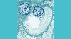
The defining feature of TBMN is diffuse thinning of the GBM, typically to less than 200-250 nm, observed on electron microscopy. In contrast to other glomerular diseases, there is an absence of significant immune deposits or inflammatory changes. This distinguishes TBMN from IgAN, which involves IgA-dominant immune deposits, and MPGN, characterized by double-contoured basement membranes.
Differentiating TBMN from Alport syndrome is critical. Although both conditions exhibit GBM thinning, Alport syndrome is associated with progressive renal dysfunction, sensorineural hearing loss, and ocular abnormalities, features absent in TBMN. Genetic testing for mutations in COL4A3 or COL4A4 can aid in distinguishing TBMN from early-stage Alport syndrome, because these mutations may be shared.
TBMN is generally considered a benign condition with an excellent prognosis. Most patients require no specific treatment beyond regular monitoring of renal function and blood pressure. Unlike IgAN or MPGN, which may necessitate immunosuppressive therapy or nephroprotective agents such as ACE inhibitors, TBMN typically does not progress to chronic kidney disease or require such interventions.
5. Alport syndrome is distinguished from IgAN and TBMN by its progressive nature, systemic manifestations, and specific GBM ultrastructural changes.
Alport syndrome is a hereditary disorder caused by mutations in the COL4A3, COL4A4, or COL4A5 genes, which encode type IV collagen, a critical component of the GBM. The disease results in progressive GBM abnormalities, leading to hematuria, proteinuria, and eventual renal failure. Alport syndrome is also associated with sensorineural hearing loss and ocular abnormalities, distinguishing it from primary glomerular diseases such as IgAN and TBMN.
The hallmark of Alport syndrome is persistent hematuria, often detected in childhood, and progressive renal dysfunction. Extrarenal features include bilateral high-frequency sensorineural hearing loss, anterior lenticonus, and retinal flecks. These systemic manifestations are absent in IgAN, which typically presents with episodic gross hematuria or persistent microscopic hematuria, often triggered by infections.
In Alport syndrome, electron microscopy reveals irregular thickening, thinning, or lamellation (“basket-weave” appearance) of the GBM. In contrast, IgAN shows mesangial IgA deposits and TBMN is characterized solely by uniformly thin GBMs without structural disarray. Genetic testing for COL4 mutations confirms Alport syndrome, aiding differentiation from TBMN, which may share similar genetic mutations in milder forms.
Whereas IgAN management includes blood pressure control and immunosuppressants in severe cases, treatment for Alport syndrome focuses on delaying renal progression with ACE inhibitors or ARBs. Unlike TBMN, which rarely progresses, Alport syndrome frequently leads to end-stage renal disease, necessitating dialysis or transplantation. Early recognition and genetic confirmation are vital to guide appropriate interventions and counseling for affected families.
Editor's Note: This article was created using several editorial tools, including AI, as part of the process. Human editors reviewed this content before publication.
Credits:
Lead Image: Nemes Laszlo/Science Source
Medscape Nephrology © 2025 WebMD, LLC
Any views expressed above are the author's own and do not necessarily reflect the views of WebMD or Medscape.
Cite this: Arnold B. Alper. Immunoglobulin A Nephropathy (IgAN): 5 Differential Diagnoses to Know - Medscape - Jan 23, 2025.






Comments