Thank you for visiting nature.com. You are using a browser version with limited support for CSS. To obtain
the best experience, we recommend you use a more up to date browser (or turn off compatibility mode in
Internet Explorer). In the meantime, to ensure continued support, we are displaying the site without styles
and JavaScript.
Pericyte KATP hyperactivity drives deep cortical hypoperfusion
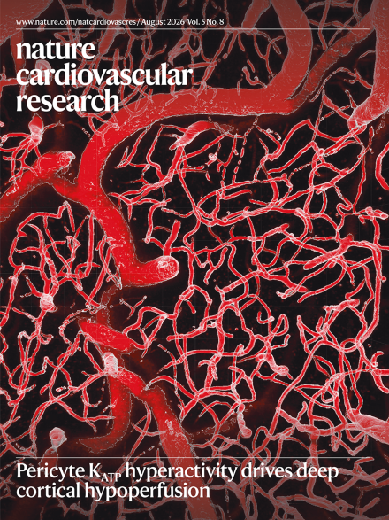
The image represents the acute cranial window imaging of the mouse cerebral vasculature. After TRITC–dextran injection, the vascular lumen was visualized in red using two-photon microscopy. Three-dimensional reconstructions of the vascular network were generated and rendered in Imaris (white) to enable detailed assessment of changes in vessel diameter.
Microfluidic, organoid, and in silico platforms have transformed our ability to investigate thrombosis-related biology, but preclinical in vivo models remain uniquely able to capture the interactions that shape thrombotic disease. Nevertheless, these models are not interchangeable and need to be carefully selected.
Myocardial infarction can cause lasting cardiac damage, heart failure and death, and immune responses are increasingly recognized as key determinants of outcomes. A study now shows that ST-segment elevation myocardial infarction (STEMI) mobilizes immature neutrophil progenitors from the bone marrow, predicting increased mortality risk.
Cerebral hemodynamic dysfunction drives unhealthy brain aging, but the role of capillaries remains unclear. Using spatial transcriptomic and advanced imaging, a study now reveals how pericyte dysfunction disrupts blood-flow distribution, causing deep-brain hypoperfusion despite preserved overall cerebral blood flow.
In a mouse model, reducing ventricular workload promotes adult cardiomyocyte proliferation through NRG1–ERBB4 signaling between epicardial cells and cardiomyocytes; this signaling activates the pentose phosphate pathway to provide metabolic support.
Santin et al. review the emerging role of lysosomes as central regulators of cardiac aging, highlighting their functions in coordinating metabolism, signaling and cellular homeostasis. They discuss how lysosomal dysfunction contributes to age-related decline and outline lysosome-targeted strategies as promising approaches to preserve cardiac function and resilience.
Richter et al. show that the mobilization of neutrophil progenitors assessed from routine blood tests can predict 30-day mortality in patients following ST-elevation myocardial infarction and enable rapid risk stratification.
Jiang et al. show that mechanical unloading promotes cardiomyocyte proliferation by enhancing epicardial–cardiomyocyte communication through the NRG1–ERBB4 axis and STAT3 activation.
By integrating spatial transcriptomics, advanced in vivo imaging, ex vivo myography, electrophysiology, in vitro functional assays and in silico modeling, Jeffrey et al. delineate a previously unrecognized mechanism of cerebral blood flow dysregulation and highlight the contribution of microvascular compartments to unhealthy brain aging.
Adusumalli et al. used spatial transcriptomics of human pluripotent stem-cell-derived cardiac progenitor grafts transplanted into pig hearts to identify Midkine as a regulator of neovascularization.
Fathieh et al. investigate the clinical value of a pre-screening strategy based on high-sensitivity troponin levels to identify patients at risk of coronary artery disease and in need of imaging diagnosis, even in the absence of traditional risk factors.