. 2025 Jul 25;11(30):eadu6990.
doi: 10.1126/sciadv.adu6990. Epub 2025 Jul 23.

A fossilized ventral ganglion reveals a chaetognath affinity for Cambrian nectocaridids

Affiliations

A fossilized ventral ganglion reveals a chaetognath affinity for Cambrian nectocaridids

Jakob Vinther et al. Sci Adv. .

Abstract

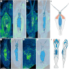
Nectocaridids are enigmatic Palaeozoic animals with a controversial phylogenetic position. Previous hypotheses have placed them in their own phylum, chordates, molluscs (specifically cephalopods), or radiodont panarthropods. We describe here a nectocaridid, Nektognathus evasmithae gen. et sp. nov. from the early Cambrian (~519 million years) Sirius Passet Lagerstätte of North Greenland. Key specimens preserve paired, phosphatized arcuate structures consistent with preservation of a ventral ganglion, a feature characteristic of extant and fossil chaetognaths, including the amiskwiid Timorebestia koprii also from Sirius Passet. Nektognathus shares a gnathostomulid-like jaw apparatus, lateral fins, subterminal anus, and large antennae with Timorebestia and Amiskwia, placing nectocaridids in the chaetognath stem lineage. The complex sensory anatomy of nectocaridids, which is partially shared with other extinct amiskwiids, highlights a more dynamic predatory lifestyle much higher in the trophic food chain during early chaetognath evolution.


Figures

Fig. 1.
Fig. 1.. Nektognathus evasmithae gen et sp. nov. holotype, MGUH34956.
(A) Image with low-angle lighting illuminated from top left, coated with magnesium oxide smoke. (B) Illuminated submerged in water under high-angle illumination. (C) EPMA carbon elemental map. (D) Interpretative drawing. (E to G) Close-up of anterior trunk region preserving the jaw apparatus. (E) Carbon elemental map featuring the outline of the jaw apparatus due to splitting of the specimen. (F) Low-angle lighting illumination coated with magnesium smoke. (G) Low-angle illumination coated with magnesium smoke with outline of jaw apparatus superimposed. (H and I) Specimen of Isoxys in the anterior section of the digestive tract, outlined in (I). (J and K) Preservation of ventral ganglion with superimposed striated musculature outlined in (K). (L and M) Digestive tract preserved by modest relief in the posterior trunk and caudal region, outlined in (M). Colors in interpretative drawing represent gut (green), ventral ganglion and nervous system (blue), jaw apparatus (red and mustard yellow), eyes (light yellow), Isoxys specimen (pink) and body outline (gray). a, antennae; cr, caudal region; fr, fin rays.
Fig. 2.
Fig. 2.. N. evasmithae gen et sp. nov. additional specimens.
Each specimen illuminated in high-angle lighting submerged in water on the left. Image is a high dynamic range (HDR) image made from several images with different high-angle illuminations. Each right-hand panel is an EPMA carbon map. (A and B) MGUH34957, smallest studied individual. (C and D) MGUH34958, small individual. (E and F) MGUH34959. (G and H) MGUH34960. (I and J) MGUH34961, EPMA carbon map in (J) is a composite with anterior region from counterpart superimposed on to the EPMA map from the main part, outlined with a stippled line. (K and L) MGUH34962.
Fig. 3.
Fig. 3.. Jaw apparatus in N. evasmithae gen. et sp. nov.
(A to E) MGUH34962. (A) Overview image, low-angle illumination from upper left and coated with magnesium oxide, and with subsequent panels denoted with squares. (B) Right lateral jaw element, outlined in (C). (D) Basal plate, outlined in (E). (F to J) MGUH34963. (F) Overview image, low-angle illumination from upper left and coated with magnesium oxide, and with subsequent panels denoted with squares. (G) Left lateral jaw element, outlined in (H). (I) Basal plate, outlined in (J). (K) Schematic outline of N. evasmithae gen. et sp. nov. with the proportions of the jaw apparatus relative to overall body dimensions highlighted. Colors: Lateral jaw elements (mustard yellow) and basal plate (red).
Fig. 4.
Fig. 4.. Phosphatized ventral ganglion in N. evasmithae gen. et sp. nov.
(A) MGUH34964, very large individual, imaged with polynomial texture mapping (PTM) specular enhancement (low angle from NW). Ventral ganglion highlighted in blue. (B and C) Close-up of ventral ganglion in counterpart imaged in low-angle illumination and with magnesium oxide smoke. (D) MGUH34960 imaged with PTM specular enhancement. Ventral ganglion highlighted in blue. (E and F) Close-up of ventral ganglion, imaged with PTM specular enhancement. (G) Extant chaetognath, sagittid sp. that have been histochemically labeled for nuclei [blue, previously figured (28)] highlighting the presence of the lateral neuron somata of the ventral ganglion enriched in nuclei, giving rise to phosphatization of ventral ganglia in total group chaetognaths in Sirius Passet (28).
Fig. 5.
Fig. 5.. Nervous system in N. evasmithae gen. et sp. nov.
(A and B) MGUH34956 holotype. (A) EPMA carbon map. (B) Outline of preserved nervous system. (C and D) MGUH34960. (C) EPMA carbon map. (D) Outline of preserved nervous system. (E) Schematic outline of possible nervous system trajectory based on available material. (F and G) MGUH34957. (F) EPMA carbon map. (G) Outline of preserved nervous system. (H and I) MGUH34962. (H) EPMA carbon map. (I) Outline of preserved nervous system. (J) Schematic nervous system in living chaetognaths, modified from Figure 2a and b in Harzsch and Müller (34) under CC BY 2.0 license (https://creativecommons.org/licenses/by/2.0/).
Fig. 6.
Fig. 6.. Jaw apparatus in Nectocaris from Chengjiang and Burgess shale.
(A to F) Nectocaris latus HKMF-50001 illuminated dry with oblique lighting. (A) Close-up of anterior and medial portion of specimen. (B) Close-up of anterior region preserving jaw apparatus, lateral jaw elements, and basal plate highlighted in (C). [(D) to (F)] N. latus ANSK-20001 illuminated dry with oblique lighting. (D) Close-up of anterior and medial portion of specimen. (E) Close-up of anterior region preserving jaw apparatus, lateral jaw elements highlighted in (F). (G to I), N. pteryx ROM 59660 (counterpart) illuminated with oblique lighting while dry. (G) Overview of entire specimen. (H) Close-up of jaw apparatus in anterior region, highlighted in (I). (J to N) N. pteryx ROM 59661 counterpart, laterally compressed specimen illuminated while submerged in water and using high-angle cross-polarized lighting. (J) Closeup of anterior and medial region of specimen. (K) Jaw apparatus highlighted in close-up view. (L) Close-up of anterior region, the same view as in (K). (M) Close-up of putative basal plate preserved in laterally compacted view, highlighted in (N). (O and P) Schematic outline of Nectocaris pteryx and N. latus with the proportions of the jaw apparatus relative to overall body dimensions highlighted. Colors: Lateral jaw elements (mustard yellow) and basal plate (red). Images of N. latus from (22). jw, jaw; bp, basal plate.
Fig. 7.
Fig. 7.. Phylogenetic analysis.
(A) Majority (50%) rule consensus tree from a Bayesian analysis (228 characters and 58 taxa), placing nectocaridids on the chaetognath stem as a sister taxon to Timorebestia (support low, 56). (B) Schematic summary phylogeny with major body plans shown and the position of the jaw apparatus highlighted.
Fig. 8.
Fig. 8.. Reconstruction of N. evasmithae gen. et sp. nov.
(A) Oblique ventral view. (B) Lateral view. (C) Dorsal view. (D) Ventral view, musculature removed. (E) Ventral view.

Similar articles

References

    1. Conway Morris S., The fossil record and the early evolution of the Metazoa. Nature 361, 219–225 (1993).
    1. Budd G. E., Jensen S., A critical reappraisal of the fossil record of the bilaterian phyla. Biol. Rev. 75, 253–295 (2000). - PubMed
    1. Briggs D. E. G., Fortey R. A., Wonderful strife: Systematics, Stem groups, and the phylogenetic signal of the Cambrian radiation. Paleobiology 31, 94–112 (2005).
    1. J. Valentine, H. Hamilton, “Body plans, phyla and arthropods,” in Arthropod Relationships (Springer, 1998), pp. 1–9.
    1. S. J. Gould, Wonderful Life: The Burgess Shale and the Nature of History (WW Norton & Company, 1989).

LinkOut - more resources